Transmission Electron Microscopy
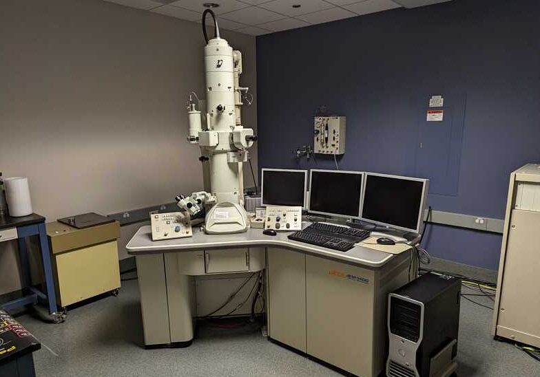
From one sample to 101, we’re here to image your answers. There’s practically no tissue we haven’t worked with.
From the more routine hepatotoxicity and phospholipodosis questions to more focused investigations of Bruch’s membrane, if it can be put on a grid, we’ll image it.
We do non-tissue samples, too! Negative staining of capsids, botanical samples, polymers….whatever goes!
Tissue processing – Resin Embedding
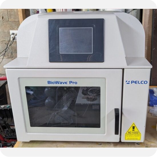
Microwave-assisted or conventional, Epon-blends or LRWhite, we’ve got you covered.
Or enrobed.
“Large” samples? Sure.
Non-Traditional samples. Yup.
Toluidine Blue stained nerves for G-Radio calculations? Done.
Whole Slide Imaging (WSI) / Slide Scanning
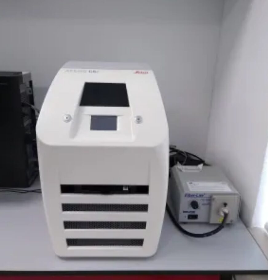
Pathology isn’t just glass anymore; it’s pixels and LUT’s, too. Whole-slide imaging is the name of the 2020’s game. From speeding consultation of areas of interest for TEM, scanning resin embedded tissue sections for analysis, to scanning your slides in our facility, we’ll take care of it. eSlides are available for viewing and/or downloading from our secure in-house server in .svs format.